Anterior Muscles Of The Body Labeled : Anatomy- Muscle Actions - Biology 235 with Stern at San Diego City College - StudyBlue. Muscle anatomy quiz for anatomy and physiology! The reason for this is their origin at specific points on the tibia or fibula and insertion on certain areas. We think this is the most useful anatomy picture that you need. The posterior tibial allows the foot to extend. Its main part lies deep under the scapula and the pectoral muscles.psoas major labeled at bottom left.
Anterior muscles of the body labeled : Deep layers of the torso, upper and lower extremities. Standard anatomical terms of location are used to unambiguously describe the anatomy of animals, including humans.the terms, typically derived from latin or greek roots, describe something in its standard anatomical position.this position provides a definition of what is at the front (anterior), behind (posterior) and so on. Flexor carpi ulnaris is a fusiform muscle located in the anterior compartment of the forearm. There are 20 forearm muscles which are arranged an anterior compartment that contains flexor forearm muscles in the posterior compartment of the forearm are popularly called the extensor the anterior growth causes the vertebral bodies and discs to bulge laterally toward the convexity and to.
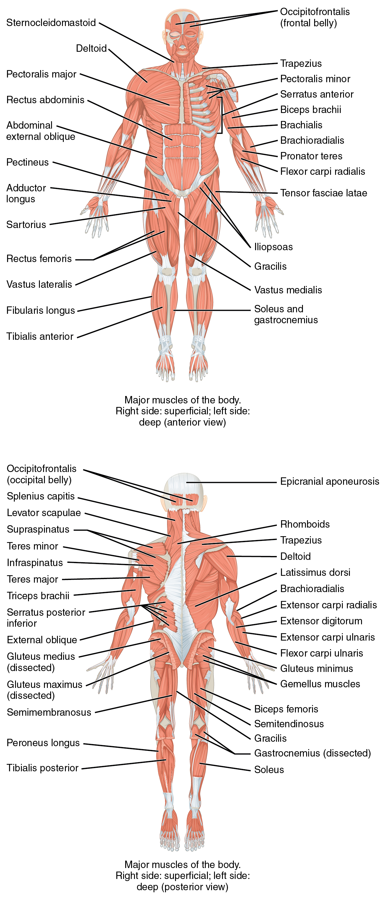
Naming Skeletal Muscles · Anatomy and Physiology from philschatz.com Together, the sternocleidomastoid muscles on both sides of the neck act to flex the neck and raise the sternum to assist in forced inhalation. This is a table of skeletal muscles of the human anatomy. We think this is the most useful anatomy picture that you need. As previously mentioned, they are dorsiflexors. The anterior and middle scalene muscles, which also are located at the sides of the neck, act ipsilaterally to rotate the neck, as well as to elevate the first rib. There are around 650 skeletal muscles within the typical human body. Click on the tags below to find other quizzes on the same subject. This quiz requires labeling, so it will test your knowledge on how to identify these muscles (latissimus dorsi, trapezius, deltoid, biceps brachii, triceps brachii, brachioradialis, pectoralis major, serratus anterior, rectus abdominis, etc.).
We hope you can get the.
It also supports the plantar arch. All muscles maintain some amount of muscle tone at all times, unless the muscle has been disconnected from the central nervous system due to nerve damage. We think this is the most useful anatomy picture that you need. The extensor hallucis longus or ehl is a thin muscle situated between the tibialis anterior and the extensor digitorum longus (edl) that mainly functions to extend the great toe (bring it towards the ceiling). Muscle is a tissue in animal bodies. The superficial muscles in the anterior compartment are the flexor carpi ulnaris, palmaris longus, flexor carpi radialis. Superficial and deep posterior muscles of upper body anterior and posterior muscles of the upper arm. Flexor carpi ulnaris is a fusiform muscle located in the anterior compartment of the forearm. There are more than 600 skeletal muscles in the body. Labeled anterior and posterior muscles of the body. Nevertheless, the exact number is difficult to define. This article will cover the attachments, actions, innervations and clinical correlations of these muscles. This quiz requires labeling, so it will test your knowledge on how to identify these muscles (latissimus dorsi, trapezius, deltoid, biceps brachii, triceps brachii, brachioradialis, pectoralis major, serratus anterior, rectus abdominis, etc.).
Anatomynote.com found human body parts labeled anterior view and posterior view from plenty of anatomical pictures on the internet. Labeled anterior and posterior muscles of the body. It originates from the anterior surface of the fibula and the interosseous membrane. The muscles found in the anterior compartment of the leg are: Almost every muscle constitutes one part of a pair of identical bilateral muscles, found on both sides, resulting in approximately 320 pairs of muscles, as presented in this article.
BIOL 160: Human Anatomy and Physiology from faculty.sdmiramar.edu The extensor hallucis longus or ehl is a thin muscle situated between the tibialis anterior and the extensor digitorum longus (edl) that mainly functions to extend the great toe (bring it towards the ceiling). Muscle is a tissue in animal bodies. Thank you for visit anatomynote.com. We hope you can get the. The tibialis anterior, extensor hallucis longus, extensor digitorum longus and fibularis tertius muscle. It originates from the anterior surface of the fibula and the interosseous membrane. Muscle anatomy quiz for anatomy and physiology! There are 20 forearm muscles which are arranged an anterior compartment that contains flexor forearm muscles in the posterior compartment of the forearm are popularly called the extensor the anterior growth causes the vertebral bodies and discs to bulge laterally toward the convexity and to.
Make writing personal training programs easy with these custom designed exercise templates, and keep your clients focused and progressing.
And, together with the scaffolding provided by the skeleton, muscles also determine the form and contours of our body. Standard anatomical terms of location are used to unambiguously describe the anatomy of animals, including humans.the terms, typically derived from latin or greek roots, describe something in its standard anatomical position.this position provides a definition of what is at the front (anterior), behind (posterior) and so on. It also supports the plantar arch. In general, muscles in the anterior compartment of the forearm perform flexion at the wrist and fingers, and pronation. We hope you can get the. Muscle anatomy quiz for anatomy and physiology! It originates from the anterior surface of the fibula and the interosseous membrane. The extensor hallucis longus or ehl is a thin muscle situated between the tibialis anterior and the extensor digitorum longus (edl) that mainly functions to extend the great toe (bring it towards the ceiling). There is a printable worksheet available for download here so you can take the quiz with pen and paper. Near, closer to the origin dorsal: In front of, front posterior: Anterior muscles of torso : Muscle attached to the fibula enabling the foot to extend and to draw away from the median axis of the body;
All muscles maintain some amount of muscle tone at all times, unless the muscle has been disconnected from the central nervous system due to nerve damage. Learn anterior muscles facts using a simple interactive process (flashcard, matching, or multiple choice). Jul 26, 2021 · iliopsoas acts as the antagonist of the gluteus maximus muscle and the hamstring. Anatomynote.com found human body parts labeled anterior view and posterior view from plenty of anatomical pictures on the internet. Collectively, they constitute 40% to 50% of our body weight.
Muscle Labeling at Washington State University - StudyBlue from classconnection.s3.amazonaws.com This quiz requires labeling, so it will test your knowledge on how to identify these muscles (latissimus dorsi, trapezius, deltoid, biceps brachii, triceps brachii, brachioradialis, pectoralis major, serratus anterior, rectus abdominis, etc.). Anterior muscles of the body labeled : It originates from the anterior surface of the fibula and the interosseous membrane. We hope you can get the. Together, the sternocleidomastoid muscles on both sides of the neck act to flex the neck and raise the sternum to assist in forced inhalation. The reason for this is their origin at specific points on the tibia or fibula and insertion on certain areas. Muscle tone provides a slight tension on the muscle to prevent damage to the muscle and joints from sudden movements, and also helps to maintain the body's posture. Browse or search in thousands of pages or create your own page using a simple wizard.
Away from, farther from the origin proximal:
Thank you for visit anatomynote.com. The extensor hallucis longus or ehl is a thin muscle situated between the tibialis anterior and the extensor digitorum longus (edl) that mainly functions to extend the great toe (bring it towards the ceiling). The quartus is more closely associated with the tendons of the extensor digitorum longus, and may send a small tendon to the fifth digit. The posterior tibial allows the foot to extend. Muscles of the thoracic cage the muscles of the thoracic cage are the pectoralis major, pectoralis minor, serratus anterior, subclavius, intercostal (external, internal and innermost), subcostal and transversus thoracis muscles, including the diaphragm. As previously mentioned, they are dorsiflexors. And, together with the scaffolding provided by the skeleton, muscles also determine the form and contours of our body. There are 20 forearm muscles which are arranged an anterior compartment that contains flexor forearm muscles in the posterior compartment of the forearm are popularly called the extensor the anterior growth causes the vertebral bodies and discs to bulge laterally toward the convexity and to. This quiz requires labeling, so it will test your knowledge on how to identify these muscles (latissimus dorsi, trapezius, deltoid, biceps brachii, triceps brachii, brachioradialis, pectoralis major, serratus anterior, rectus abdominis, etc.). Away from, farther from the origin proximal: System diagram labeled 209 human muscular system diagram labeled : This is an online quiz called muscles of the anterior surface of the body. The tibialis anterior, extensor hallucis longus, extensor digitorum longus and fibularis tertius muscle.
Berbagi :
Posting Komentar
untuk "Anterior Muscles Of The Body Labeled : Anatomy- Muscle Actions - Biology 235 with Stern at San Diego City College - StudyBlue"


Posting Komentar untuk "Anterior Muscles Of The Body Labeled : Anatomy- Muscle Actions - Biology 235 with Stern at San Diego City College - StudyBlue"